Malondialdehyde (MDA) Content Assay Kit
Note: It is necessary to predict 2-3 large difference samples before the formal determination. Operation Equipment: Spectrophotometer.
Cat No: BC0020
Size: 50T/48S
Components:
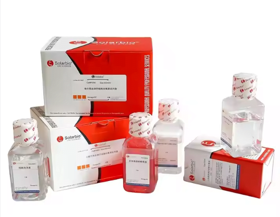
| Component | Type | Volume/Quantity | Storage |
|---|---|---|---|
| Extraction reagent | Liquid | 60 mL × 1 | 4°C |
| Reagent I | Liquid | 42 mL × 1 | 4°C |
| Reagent II | Powder | – | 4°C |
| MDA working reagent | Liquid | 20 mL Reagent I | 4°C |
| + Reagent II | |||
| Reagent III | Liquid | 12 mL × 1 | 4°C |
Note: The working solution for MDA detection is difficult to dissolve, which can be heated at 70℃ and vibrated violently to promote dissolution. Or by ultrasonic treatment to promote dissolution.
Product Description:
- Lipid peroxide is generated through the interaction of oxygen free radicals with unsaturated fatty acids, eventually forming compounds such as malondialdehyde (MDA). The level of lipid peroxidation serves as an indicator, which can be assessed by measuring MDA levels.
- Under acidic and high-temperature conditions, a brown-red compound, 3,5,5-three methyl sulfamethoxazole-2,4-two ketone, is synthesized via a condensation reaction between MDA and thiobarbituric acid (TBA). This compound exhibits maximum absorption at 532 nm. The assessment of lipid peroxidation involves colorimetric analysis. However, the presence of soluble sugars can interfere with the detection process. The reaction of soluble sugars with TBA produces a color reaction with absorption wavelengths at 450 nm and 532 nm. In this assay kit, the MDA content is determined by the variance in absorbance at 532 nm, 450 nm, and 600 nm.
- Due to the presence of sucrose in plant tissues and glucose in animal tissues, the kit includes two computational formulas tailored for sucrose and glucose. These formulas are suitable for lipid assessment.
Reagents and Equipment Required but Not Provided:
Spectrophotometer, water bath, desk centrifuge, transfer pipette,1 mL glass cuvette, mortar/homogenizer, ice and distilled water.
Procedure:
I. Sample preparation:
Bacteria or cells:
Collect bacteria or cells into the centrifuge tube. 5 million bacteria or cells could be mixed with 1 mL of Extraction reagent. Use ultrasonication to split bacteria and cells (placed on ice, ultrasonic power 200W, ultrasonic time 3 seconds, interval 10 seconds, repeat for 30 times). Centrifuge at 8000 ×g for 10 minutes at 4℃ to remove insoluble materials, and take supernatant on ice before testing.
Tissue:
0.1 g of tissue could be mixed with 1 mL of Extraction reagent and fully homogenized on ice bath. Then centrifuge at 8000 ×g for 10 minutes at 4℃ to remove insoluble materials, and take the supernatant on ice before testing.
Serum:
Detect
II. Determination procedure:
- Preheat spectrophotometer for more than 30 minutes, set zero with distilled
- Add reagents with the following list:
| Reagent (μL) | Test tube (T) | Blank tube (B) |
| MDA working reagent | 600 | 600 |
| Sample | 200 | – |
| Distilled water | – | 200 |
| Reagent Ⅲ | 200 | 200 |
The mixture would be incubated at 100℃ for 60 minutes (tightly close to prevent moisture loss), cooled on ice, and centrifuged at 10000 ×g for 10 minutes at room temperature to remove insoluble materials. Take supernatant in 1 mL glass cuvette, and measure the absorbance at 450 nm, 532 nm and 600 nm.
∆A450=A450(T)-A450(B), ∆A532=A532(T)-A532(B), ∆A600 =A600(T)-A600(B). Blank tube needs to test once or twice.
III. Calculation:
- Tissue, bacteria or cultured cells
- Protein concentration:
MDA (nmol/mg prot)=(6.45×(∆A532-∆A600)-1.29×∆A450)×Vrv÷(Cpr×Vs)
=5×(6.45×(∆A532-∆A600)-1.29×∆A450)÷Cpr
- Sample weight:
MDA (nmol/g weight)=( 6.45×(∆A532-∆A600)- 1.29×∆A450)×Vrv÷(W×Vs÷Vsv)
=5×(6.45×(∆A532-∆A600)- 1.29×∆A450)÷W
- Cellamount:
MDA (nmol/104cell)=( 6.45 ×(∆A532-∆A600)- 1.29×A∆450)×Vrv÷(400×Vs÷Vsv)
=0.01×(6.45×(∆A532-∆A600)- 1.29×∆A450)
- Serum:
MDA (nmol/mL)=(6.45×(ΔA532 -ΔA600)-1.29×ΔA450)×Vrv÷Vs
=5×(6.45×(ΔA532 -ΔA600)-1.29×ΔA450)
- Plants tissue:
- Sample weight
MDA (nmol/g weight)= (6.45×(∆A532-∆A600)-0.56×∆A450)×Vrv÷(W×Vs÷Vsv)
=5×(6.45 ×(∆A532-∆A600)- 0.56×∆A450)÷W
- Protein concentration:
MDA (nmol/mg prot)=( 6.45 ×(∆A532-∆A600)- 0.56×∆A450)×Vrv÷(Cpr×Vs)
=5×(6.45 ×(∆A532-∆A600)- 0.56×∆A450)÷Cpr
Vrv: Total reaction volume, 1 mL; Vs: Sample volume, 0.2 mL;
Vsv: Extraction volume, 1 mL;
Cpr: Sample protein concentration, mg/mL; W: Sample weight, g;
400: Total number of bacteria and cells, 5 million.
Note:
If it is found that the absorbance value of the sample is too low, the boiling water bath time can be adjusted from 60 minutes to 90 minutes or longer. The detection of MDA in the same experiment needs to be extended to the same time to avoid errors.
References
- Spitz D R, Oberley L W. An assay for superoxide dismutase activity in mammaliantissue homogenates[J]. Analytical Biochemistry,1989
- Masayasu M, Hiroshi Y. A simplified assay method of superoxide dismutase activity for clinical use[J]. Clinica Chimica
Related products:
BC3590/BC3595 Hydrogen Peroxide (H2O2) Content Assay Kit
BC1090/BC1095 Xanthine Oxidase (XOD) Activity Assay Kit
BC0690/BC0695 Glucose Oxidase (GOD) Activity Assay Kit BC1270/BC1275 Protein Carbonyl Content Assay Kit BC1280/BC1285 Diamine Oxidase (DAO) Activity Assay Kit BC1290/BC1295 Superoxide Anion Content Assay Kit
Customers’ Reviews of Solarbio Products for Reference
Reviews
There are no reviews yet.