With the development of molecular biology techniques, various methods for detecting gene structures and mutations have continuously emerged. Especially after the advent of PCR technology, various gene detection techniques combined with PCR have further propelled the advancement of gene research. Techniques such as direct sequencing of asymmetric PCR products, ribonuclease cleavage (RNAase), and restriction fragment length polymorphism analysis (RFLP) have become powerful tools for gene analysis. However, these methods are relatively cumbersome to operate, have significant limitations, or require high experimental conditions, making them unsuitable for general clinical laboratories.
Introduced in 1989, PCR-SSCP (Single-Strand Conformation Polymorphism) has undergone continuous improvement and refinement, making it a simpler, faster, and more sensitive method for detecting gene mutations. It is not only used for detecting point mutations and short sequence deletions and insertions but is also applied in DNA quantification, monitoring cross-contamination in PCR diagnostic experiments, and investigating sources of infection. Due to the outstanding advantages of SSCP, it has been widely applied in recent years.

Principles and Characteristics of SSCP
- Discovery and Basic Concept:
- Single-stranded DNA (ssDNA) fragments exhibit complex spatial folding conformations maintained by intramolecular interactions, primarily base pairing.
- A single base change can significantly or slightly alter the spatial conformation, resulting in different migration patterns in polyacrylamide gel.
- Separation Mechanism:
- Non-denaturing polyacrylamide gel electrophoresis (PAGE) can sharply separate ssDNA molecules with different conformations due to varying levels of obstruction in the gel.
- SSCP Analysis:
- Named Single-Strand Conformation Polymorphism (SSCP) by Japanese researcher Orita and colleagues.
- Applied to detect gene mutations in PCR-amplified products, enhancing simplicity and sensitivity.
- Basic Process:
- PCR Amplification: Amplify the target DNA.
- Denaturation and Renaturation: Denature the specific PCR products and rapidly renature them to form single-stranded DNA molecules with specific spatial structures.
- Non-denaturing PAGE: Perform non-denaturing polyacrylamide gel electrophoresis on an appropriate amount of single-stranded DNA.
- Detection: Analyze the results using radiographic autoradiography, silver staining, or ethidium bromide staining.
- Interpretation:
- A change in the migration rate of ssDNA compared to the normal control indicates a change in conformation, suggesting a base mutation in that DNA fragment.
- Advantages:
- Simple, fast, and sensitive method.
- Does not require specialized equipment.
- Suitable for clinical laboratories.
- Limitations:
- Only detects mutations; further sequencing is needed to pinpoint the exact position and type of mutation.
- Strict electrophoresis conditions required.
- Possible false negatives if point mutations do not significantly alter the ssDNA conformation or if other conditions interfere.
- Detection Efficiency:
- Despite limitations, SSCP boasts a high detection rate compared to other methods.
- Capable of identifying unknown base mutations within the target DNA fragment.
- Takao demonstrated that SSCP can detect 90% of single-base mutations in DNA fragments shorter than 300 bp.
- Further Applications:
- SSCP can separate mutant ssDNA with different migration rates through polyacrylamide gel electrophoresis.
- Allows for further purification and identification of mutant DNA fragments at the sequence level.
Development and Improvements of SSCP
- Initial Development:
- Initially, SSCP involved incorporating isotopes into PCR amplification products, and results were displayed through autoradiography. This posed challenges for widespread adoption.
- Simplification:
- The combination of DNA silver staining with PCR-SSCP, particularly the application of direct ethidium bromide staining, greatly simplified the method.
- Recent Noteworthy Improvements:
- RNA-SSCP Analysis:
- The basic principle: RNA has more intricate secondary and tertiary conformations, making it highly sensitive to single-base mutations, thus increasing detection rates.
- Mutation detection rate can exceed 90%.
- RNA is less prone to forming double strands, allowing for a larger quantity to be used in electrophoresis, which is beneficial for ethidium bromide staining.
- However, this method adds a reverse transcription step and requires longer primers containing RNA polymerase promoter sequences, increasing complexity.
- RNA-SSCP Analysis:
- Combining SSCP with Other Mutation Detection Methods:
- Heteroduplex Analysis (Het):
- Improves detection rates significantly when combined with SSCP.
- Involves hybridizing a probe with the target ssDNA or RNA. Hybrid strands containing a mismatched base pair can be separated from completely complementary hybrid strands through electrophoresis in non-denaturing PAG gel.
- Performing both SSCP and Het analysis on the same target sequence can achieve near 100% detection rate for point mutations, while maintaining experimental simplicity.
- Heteroduplex Analysis (Het):
The Procedure of PCR-SSCP
Gel Preparation Based on DNA Fragment Length
For DNA fragments shorter than 1Kb, choose the concentration of polyacrylamide gel as follows:
- DNA Fragment Length (bp): % Acrylamide Concentration
- 1Kb–700b: 3.5%
- 700b–500b: 5%
- 500b–200b: 8%
- 200b: 12%
Pre-SSCP Preparations
- PCR Amplification: Amplify a specific product with minimal smearing (confirmed by agarose gel electrophoresis).
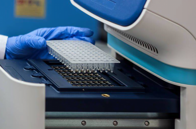
1. Preparation of Polyacrylamide Gel (PAG)
- Prepare the gel solution based on the required concentration from the table above.
- Insert a comb into the gel mold.
- Pour the gel solution from one end of the comb, tilting the mold towards the pouring end as the gel reaches the comb teeth to prevent bubble formation.
- Let the gel solidify at room temperature for 1 hour.
- Remove the comb and add 1×TBE buffer on top of the gel to cover it.
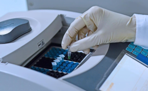
2. Electrophoresis
- Take 10 μl of PCR product.
- Add 10 μl of denaturing agent (95% formamide, 10 mmol/L EDTA, 0.02% bromophenol blue).
- Add 30 μl of mineral oil.
- Boil for 5 minutes, then immediately place in an ice bath for at least 2 minutes.
- Load the entire aqueous phase onto the gel.
- Perform electrophoresis at 10-15°C:
- Start with 300V for 5 minutes.
- Continue with 120V for 8 hours.
- After electrophoresis, stain the gel in 1×TBE buffer containing 0.5 μg/ml ethidium bromide for 30-45 minutes.
- Observe under UV light or proceed to silver staining.
3. Silver Staining
- Wash the PAG twice with deionized water.
- Fix the gel in a solution of 10% ethanol and 0.5% acetic acid for 6 minutes.
- Wash twice with deionized water.
- Immerse in 0.2% silver nitrate (AgNO3) solution for 10 minutes.
- Wash 3-5 times with deionized water.
- Develop in a solution of 1.5% sodium hydroxide (NaOH) and 0.4% formaldehyde for 7 minutes.
- Stop the development with 0.75% sodium carbonate (NaCO3).
Applications of SSCP
Since Orita and colleagues used SSCP for human DNA polymorphism analysis, the method has been extensively employed in various fields, including the detection of gene mutations associated with cancer and hereditary diseases, as well as in virus typing and contamination monitoring.
Cancer Gene Mutation Detection
- Tumor-Associated Mutations:
- SSCP is widely used to detect mutations in genes related to various cancers such as astrocytoma, brain tumors, small cell lung cancer, gastric cancer, and colorectal cancer.
- It has been applied to detect mutations in the P53 gene in these tumors and ras gene mutations in lung cancer.
- Successful Applications:
- Recently, Sugano et al. successfully used silver-stained SSCP to detect a mutation at position 12 of the c-Ki-ras2 gene, completing the electrophoresis and staining process within 2.5 hours.
Hereditary Disease Research
- SSCP is used in the study of genes involved in hereditary diseases such as:
- Cystic Fibrosis: Detection of mutations in the CFTR gene.
- Neurofibromatosis Type 1: Analysis of gene mutations.
- Familial Adenomatous Polyposis: Gene research related to this condition.
- RNA-SSCP vs. DNA-SSCP:
- Sharkar et al. used RNA-SSCP to detect gene sequences in 28 hemophilia B patients.
- Comparison with DNA-SSCP and direct gene sequencing revealed 20 base mutations in the 2.6kb Factor IX gene, with RNA-SSCP detecting 70% of these mutations compared to 35% detected by DNA-SSCP, demonstrating higher sensitivity of RNA-SSCP.
Virus Typing and Contamination Monitoring
- Virus Typing:
- SSCP is used for virus typing and monitoring contamination in PCR experiments.
- YaP used nested PCR to detect HBV samples from different regions, followed by SSCP analysis, which revealed distinct SSCP patterns for each sample, indicating regional variations in HBV DNA and eliminating the possibility of cross-contamination.
- Contamination Monitoring:
- SSCP serves as a reliable method to ensure the accuracy of PCR diagnostic results.
Pathogen Transmission Studies
- SSCP is utilized to study the transmission routes of pathogens.
Quantitative DNA Analysis
- Breast Cancer Cell Analysis:
- SSCP, combined with competitive PCR, was used for quantitative analysis of mutant P53 genes in breast cancer cells.
- The method’s advantage lies in the use of internal standards that differ from the target DNA by only one base, ensuring identical amplification conditions and thus more accurate DNA quantification.
Precautions for SSCP
SSCP is a rapid, simple, and sensitive method for detecting gene mutations. To achieve optimal results, the following precautions should be observed:
Repeatability:
- The main factors affecting the repeatability of SSCP are electrophoresis voltage and temperature. Keeping these conditions constant ensures good repeatability of SSCP patterns.
- Generally, SSCP patterns show two single-stranded DNA bands, but sometimes only one or more than three bands may appear due to similar spatial conformations between two single-stranded DNA molecules. The presence of more than three bands can indicate a mixture of wild-type and mutant DNA fragments.
Impact of Target DNA Sequence Length:
- SSCP is more effective in detecting point mutations in shorter DNA or RNA sequences than in longer ones. This might be because single-base changes in longer molecules have a smaller impact on maintaining spatial conformation.
- Some researchers believe that for DNA chains shorter than 400 bp, the length does not affect SSCP’s effectiveness. Careful selection of experimental conditions can achieve a detection rate of over 90% for point mutations in 354 bp DNA fragments.
Electrophoresis Voltage and Temperature:
- To maintain stable spatial conformations of single-stranded DNA, SSCP should be conducted at lower temperatures (typically between 4°C and 15°C). High voltage at the start (250V) for 5 minutes helps to initially separate single-stranded DNA with different conformations without significantly raising the gel temperature. This should be followed by lower voltage (around 100V) to further separate the strands.
- The exact voltage for electrophoresis should be determined based on specific experimental conditions.
Impact of Point Mutation Position:
- The position of a point mutation in DNA or RNA affects SSCP detection rates based on its role in maintaining spatial conformation, rather than its linear position on the chain.
- Mutations in the central region of DNA are generally easier to detect compared to those near the ends.
- However, even mutations in the central region may go undetected if they do not significantly alter the spatial conformation. For instance, White’s study found that only 2 out of 9 samples with point mutations at the loop of a hairpin structure were detected, indicating a detection rate of 22%.
Interpreting SSCP Results:
- SSCP separates single-stranded DNA based on spatial conformation rather than molecular weight or charge. Sometimes, the migration rates of normal and mutant chains are very close, making it hard to distinguish between them.
- Typically, electrophoresis lengths of 16-18 cm are required to accurately determine results based on the detection limit, which is the smallest discernible electrophoresis distance difference between mutant and normal DNA fragments.
- If the distance is greater than the detection limit (e.g., 3mm), a mutation is indicated. A smaller distance suggests no change. Setting a low detection limit can lead to false positives.
- Other conditions, such as the amount of PCR product loaded, the degree of PAG crosslinking, and gel concentration, should be selected based on specific experimental requirements.
Details of PCR-SSCP by Steps
Sample Preparation
- PCR Amplification and Detection:
- Perform PCR amplification using appropriate primers and select the suitable annealing temperature.
- Detect the amplification products by running them on a 2-2.5% agarose gel through horizontal electrophoresis. Load 3-5 µl of the PCR product.
- The optimal concentration of the PCR product should be around 10 ng/µl.
- Mixing PCR Product with Denaturing Buffer:
- Add 5 µl of SSCP sample buffer (denaturing buffer, same as the sequencing buffer in “Molecular Cloning”) to the bottom of a clean PCR tube.
- Add the PCR amplification product to the center of the buffer. Adjust the amount based on the visibility of the band on the agarose gel:
- If the band is bright, add 1 µl.
- If the result is excellent, add 0.5 µl.
- If the band is not very clear, add 1.5, 2, or 3 µl accordingly.
- Increase the amount of sample buffer proportionately, e.g., for 3 µl of PCR product, add 8 µl of buffer for better denaturation.
- Denaturation of PCR Product:
- Centrifuge the tube to concentrate the sample at the bottom.
- Denature the sample at 95°C for 10 minutes using a PCR machine, then immediately place the PCR product on ice for 5 minutes to prevent renaturation.
Note: Steps 3 and 4 can be performed after the fourth step of gel preparation while waiting for the gel to solidify.
Gel Preparation
- Preparing the Glass Plates:
- Rinse each pair of glass plates with tap water followed by ddH2O.
- Dry the glass plates with absorbent paper and then wipe with anhydrous ethanol.
- Allow the ethanol to evaporate for 2-3 minutes.
- Assemble the glass plates and place them into the gel casting stand, ensuring both sides and the bottom are secured.
- Tighten the assembly screws to keep the plates level. (Check for leaks using anhydrous ethanol.)
Large Gels (50% Glycerol Concentration)
| Component | 8% Gel (10ml lower gel) | 8% Gel (5ml stacking gel) | 6% Gel (10ml lower gel) | 6% Gel (5ml stacking gel) |
|---|---|---|---|---|
| 30% PAGE | 2.7ml | 1ml | ||
| 10× TBE | 1ml | 0.5ml | ||
| 50% Glycerol | 1ml | 0.5ml | ||
| ddH2O | 5ml | 3ml | ||
| APS | 70µl | 70µl | ||
| TEMED | 12µl | 8µl | ||
| Total Volume | 10ml | 5ml |
Small Gels (50% Glycerol Concentration)
| Component | 8% Gel (15ml lower gel) | 8% Gel (10ml stacking gel) | 6% Gel (15ml lower gel) | 6% Gel (10ml stacking gel) |
|---|---|---|---|---|
| 30% PAGE (4°C) | 4.05ml | 2ml | 2ml | |
| 10× TBE | 1.5ml | 1ml | 1ml | |
| 50% Glycerol | 1.5ml | 1ml | 1ml | |
| ddH2O | 7.5ml | 6ml | 6ml | |
| APS | 105µl | 70µl | 70µl | |
| TEMED | 12µl | 12µl | 12µl | |
| Total Volume | 15ml | 10ml | 10ml | 5ml |
Gel Pouring
- Mixing the Gel: After preparing the gel solution according to the provided formulation, thoroughly mix it to ensure uniformity.
- Pouring into Glass Plates: Carefully pour the prepared gel solution into the assembled glass plates. Ensure that no air bubbles are trapped during the pouring process. If any bubbles are observed, gently tilt the plates to allow the bubbles to rise to the surface. Lightly tapping the sides of the plates can help in releasing trapped air bubbles.
- Inserting the Comb: Once the gel solution level reaches approximately 0.5cm below the top edge of the glass plates, insert the comb into the gel. Ensure that there are no air bubbles trapped between the comb teeth and the gel surface. If any bubbles are present, remove the comb, release the bubbles, and reinsert the comb carefully.
- Allowing Gel Polymerization: After inserting the comb, allow the gel to polymerize by placing the plates either flat or at a slight angle (less than 10 degrees) on a level surface for approximately 30 minutes. During this time, the gel will polymerize and solidify.
Note: This is also a suitable time to proceed with the denaturation of PCR samples as described in steps 3 and 4 of the first section.
Gel Loading
- Placing the Gel:
- Remove the comb from the gel promptly after polymerization.
- Ensure even force is applied to maintain the integrity of the wells.
- Secure the gel plate onto the electrophoresis apparatus with the well side facing inward.
- Transition the gel plate onto the electrophoresis buffer slowly to avoid large air bubbles.
- Pre-Electrophoresis:
- Add 1× TBE electrophoresis buffer to the upper reservoir until the liquid level is about 1cm above the shorter edge of the glass plate.
- Ensure there is no leakage from the upper reservoir.
- Set the voltage to 140-150V for the large apparatus and 110-120V for the small apparatus.
- Pre-electrophoresis for approximately 10 minutes.
- Sample Loading:
- Sequentially add prepared samples into the wells of the gel using a microsyringe.
- Avoid loading samples into the two wells closest to the edges of each gel plate.
- Electrophoresis:
- Set the voltage to 140-150V for the large apparatus and 110-120V for the small apparatus.
- Perform electrophoresis at a temperature of 10-15°C for a minimum of 10 hours.
Staining
- Fixation:
- Remove the gel from the apparatus, mark the starting point, and immerse it in 70% ethanol for 15 minutes on a shaker.
- Recover the ethanol after fixation and rinse twice with distilled water for about 3 minutes per rinse.
- Staining:
- Stain the gel in staining solution (200ml containing 3.6% NaOH 4.2ml, 20% AgNO3 3.6ml, ammonia 2ml) for 30 minutes.
- Adjust the staining time if staining two gels simultaneously.
- Developing:
- Develop the gel in developing solution (200ml containing 1% sodium citrate 1ml, formaldehyde 100ul) until bands are clearly visible.
- Rinse three times with distilled water for 3 minutes each to stop the development.
- Alternatively, stain the gel by immersing it in 1× TBE buffer containing 0.5ug/ml ethidium bromide for 10 minutes and visualize under ultraviolet light
Formulas
10×TBE Formula:
- Tris Base: 108g
- Boric Acid: 55g
- 0.5mol/L EDTA (pH 8.0), volume adjusted to 1L
Denaturing Buffer Formula:
- 98% Deionized Formamide
- 10mmol/L EDTA (pH 8.0)
- 0.025% Xylene Cyanol FF
- 0.025% Bromophenol Blue
Notes:
- Reproducibility:
- Maintaining stable voltage and temperature during electrophoresis is the main factor affecting the reproducibility of SSCP.
- Typically, SSCP patterns show two single-stranded DNA bands, but sometimes only one or three or more bands may appear, possibly due to the presence of similar three-dimensional conformations between wild-type and mutant DNA fragments.
- Influence of Target DNA Sequence Length:
- SSCP has a higher detection rate for point mutations in short-chain DNA or RNA compared to long-chain DNA. This is likely because individual base changes in long-chain DNA and RNA molecules have a smaller effect on maintaining three-dimensional conformations.
- In the case of shorter DNA chains (below 400bp), the length of DNA does not affect the effectiveness of SSCP.
- Effects of Electrophoresis Voltage and Temperature:
- To maintain a stable three-dimensional conformation of single-stranded DNA, SSCP should be performed at a lower temperature (generally between 4°C and 15°C).
- During electrophoresis, excessive voltage can cause a temperature increase. Therefore, when performing SSCP in an electrophoresis tank without a cooling device, a higher voltage (250V) should be used initially, followed by a reduction to around 100V for electrophoresis.
- Interpretation of SSCP Results:
- In SSCP analysis, the separation of single-stranded DNA fragments is based on the size of their steric hindrance rather than molecular weight, thus unable to reflect the molecular weight.
- Result interpretation should be based on the detection limit, which refers to the minimum difference in electrophoretic distance between mutant and normal DNA fragments that can be distinguished.
- Other Considerations:
- Conditions such as the amount of PCR product loaded, the crosslinking density of polyacrylamide gel, and the concentration of the gel should be selected and determined based on specific experimental conditions.