
Introduction of Situ PCR
In scientific research, the establishment of each new technology brings forth a series of new research findings, thereby advancing various disciplines. In the field of morphological research, the introduction of the electron microscope in the 1950s brought detailed studies from the cellular level down to the subcellular level.
In the 1960s and 1970s, the widespread application of immunohistochemistry and immunocytochemistry technologies pushed observations from subcellular structures to the protein molecular level, enabling the localization of numerous active substances within cells or subcellular levels. This had a profound impact on the development of medical biology.
The 1970s saw the extensive application of molecular biology techniques in morphology, and with the advent of in situ hybridization, specific DNA or RNA sequences within tissues and cells could be localized. This advanced observations from the protein level to the gene level, thus deepening human understanding of many life phenomena at the genetic level.
In the 1980s, a powerful technique in the field of molecular biology, PCR (polymerase chain reaction), was introduced. It was quickly adopted in morphological studies, allowing for the localization and observation of specific low-copy or single-copy DNA or RNA within cells. The advent of this technology promises to bring more research findings, taking morphological studies a significant step forward.

Fundamental Principles
The basic principle of in situ PCR technology is to combine the high-efficiency amplification of PCR technology with the cellular localization of in situ hybridization. This allows for the in situ detection of specific low-copy or single-copy DNA or RNA sequences within tissue cells.
PCR technology amplifies specific target sequences guided by primers through cycles of denaturation, recocido, and primer extension under the action of DNA polymerase. The amplified target sequences (typically can be amplified by 10^6 times) are easily displayed through gel electrophoresis or Southern blot hybridization. Por lo tanto, PCR technology is highly sensitive and specific, and with the automation and stability of thermal cycling, it becomes easy to operate. Sin embargo, PCR is performed in a liquid phase, requiring cell disruption to extract nucleic acids as templates before amplification. This makes it difficult to correlate PCR results with the morphological structure of tissue cells and to determine the cell type containing the specific target sequence.
In situ PCR technology successfully combines PCR and in situ hybridization, retaining the advantages of both while compensating for their respective shortcomings. The samples for in situ PCR are usually chemically fixed to maintain good morphological structures of tissue cells. Both cell membranes and nuclear membranes have certain permeability, allowing components such as primers, ADN polimerasa, and nucleotides to enter cells or nuclei during PCR amplification. The fixed intracellular or intranuclear RNA or DNA serves as templates for in situ amplification. The amplified products are generally large molecules or intertwine with each other, making it difficult for them to diffuse through the cell membrane or within the membrane, thus being retained in situ. This way, the original intracellular low-copy or single-copy specific DNA or RNA sequences are exponentially amplified in situ, and the amplified products can be easily detected by in situ hybridization techniques.
Basic Types
Based on whether the nucleotides or primers used in the amplification reaction are labeled, in situ PCR technology can be divided into two major categories: direct and indirect methods. Además, there is the in situ reverse transcription PCR technology.
Direct In Situ PCR Technology
In direct in situ PCR technology, the amplified product directly carries a labeled molecule, es decir., using labeled adenosine triphosphate or primer fragments. When the specimen undergoes PCR amplification, the labeled molecules are incorporated into the amplified product, displaying the markers, thus making the specific DNA or RNA visible in the specimen (in situ).
Common markers include the radioactive isotope ³⁵S, biotin, and digoxigenin. These markers’ positions are visualized using autoradiography, affinity histochemistry, or immunohistochemistry methods.
Ventajas:
- Simple operation
- Short process
- Time-saving
Disadvantages:
- Lower specificity
- Prone to false positives
- Lower amplification efficiency, especially in paraffin sections
These disadvantages are prominent in paraffin sections due to potential DNA damage during processing (fixation, dehydration, embedding). Damaged DNA can be repaired using the labeled nucleotides, leading to false positives. Using labeled primers in direct in situ PCR further reduces amplification efficiency.
Indirect In Situ PCR Technology
Indirect in situ PCR technology involves the amplification of specific DNA or RNA within cells, followed by in situ hybridization with labeled probes, significantly improving specificity. This method is currently the most widely used in situ PCR technology.
Differences from Direct Method:
- Reaction system is the same as conventional PCR
- No labels on primers or adenosine triphosphate used
In indirect in situ PCR, amplification is achieved first, followed by detection of the amplified specific DNA products using in situ hybridization techniques. This method is also known as PCR in situ hybridization (PISH).
Ventajas:
- Higher specificity
- Higher amplification efficiency
Disadvantages:
- More complex operation steps compared to the direct method
In Situ Reverse Transcription PCR Technology
In situ reverse transcription PCR (In Situ RT-PCR) is a new technology that applies liquid-phase RT-PCR techniques to tissue cell specimens. Unlike RT-PCR (liquid phase), before performing in situ reverse transcription PCR, tissue specimens are treated with DNase to destroy any DNA present in the tissue, ensuring the template for amplification is synthesized cDNA from mRNA, not the original DNA in the cell. The other basic steps are similar to liquid-phase RT-PCR.
Basic Steps
The basic steps of in situ PCR technology include specimen preparation, in situ amplification (PCR), and in situ detection. The steps are detailed below, with a focus on paraffin sections as an example.
Specimen Preparation
In situ PCR technology can be applied to cell suspensions, cell smears, frozen sections, and paraffin sections. Compared to other methods, performing in situ PCR on suspended intact cells yields the best results, while paraffin sections yield the worst. Despite this, recent advancements have reported satisfactory PCR efficiency in paraffin sections.
Challenges:
- Poor thermal conduction and uneven heat convection on slides
- Adsorption of Taq DNA polymerase by glass slides
- Lack of intact cytoplasmic or nuclear membranes leading to diffuse amplification products
Fixation:
- Formalin or paraformaldehyde fixation (10% buffered formalin or 4% paraformaldehyde) works well for in situ PCR
- Optimal fixation time: 4-6 hours at 4°C
Section Thickness:
- Thicker sections yield better results due to higher target DNA content and more membrane structures, preventing product diffusion. Sin embargo, thicker sections can reduce morphological observation quality and resolution.
Slide Treatment:
- Prevent tissue detachment during PCR and in situ hybridization by treating slides with poly-L-lysine or silanization.
Proteinase Digestion
Before in situ amplification, tissue specimens need proteinase treatment. Proteinase digestion increases cell permeability, allowing reaction components to enter cells and exposing target sequences for amplification. Commonly used proteinases include proteinase K, trypsin, or pepsin. The extent of digestion should be adjusted based on tissue fixation. After digestion, ensure enzyme inactivation by heating or thorough washing, as residual enzymes can destroy Taq DNA polymerase in the PCR reaction.
Pros and Cons:
- Digestion increases permeability, aiding subsequent reactions
- It can also increase the chance of product diffusion, leading to false positives or negatives.
In Situ Amplification (PCR)
In situ amplification refers to performing the PCR reaction directly on tissue cell specimens. Its basic principle is entirely the same as that of liquid-phase PCR.
Imprimaciones
The primers used in PCR are typically 15-30 base pairs (pb) in length, and the amplified fragments are about 100-1000 pb. Shorter primers are preferable for in situ PCR. DNA extracted from paraffin sections is rarely over 400 pb, and RNA is seldom over 200 pb. Longer sequences are more prone to mismatches between primers and templates, leading to non-specific reactions.
Reaction System
The reaction system for in situ PCR is fundamentally similar to conventional liquid-phase PCR. Sin embargo, since it is conducted on fixed tissue sections, to achieve better amplification results, it is suggested that the concentrations of primers, Taq DNA polymerase, and Mg²⁺ in the reaction system should be higher than those in liquid-phase PCR. Bovine serum albumin (BSA) should be added to the reaction system to prevent the binding of Taq DNA polymerase to the glass slide, which could reduce amplification efficiency.
Ciclismo térmico
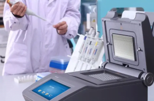
Thermal cycling for in situ PCR can be performed on a specialized thermal cycler, which is simple to operate. It can also be done on a general PCR thermal cycler by covering the sample platform with a layer of aluminum foil to create a platform, filling the space on the sample platform with mineral oil or water, and placing the slides on the platform to carry out the thermal cycling steps.
To ensure sufficient amplification, the duration of each step in the thermal cycling process for in situ PCR can be slightly longer than that for conventional PCR. Además, the hot start method can be employed, where the slide is heated to 80-94°C before immediately adding Taq DNA polymerase.
To minimize the loss of the reaction system during the thermal cycling process, clear nail polish, mineral oil, or a PAP pen can be used to seal the edges of the cover slip.
Washing
After in situ amplification, the specimen should be washed to remove the amplified products that have diffused outside the cells. Insufficient washing can result in visible amplified products during detection, causing a high background or false-positive results. Sin embargo, excessive washing may also elute the amplified products inside the cells, weakening or losing the positive signal.
Some authors recommend post-fixation with 4% paraformaldehyde for 2 hours or 2% glutaraldehyde for 5 minutes after amplification to retain the amplified products within the cells during detection, enhancing sensitivity and specificity.
In Situ Detection
The method of detecting in situ PCR amplification products depends on the in situ PCR design. The direct method involves direct in situ detection based on the nature of the labeled molecules. The indirect method requires in situ hybridization for detection.
Detection labeled with fluorescein, biotin, alkaline phosphatase, or horseradish peroxidase follows the same principles as immunohistochemistry and affinity histochemistry techniques.
Applications of In Situ PCR Technology
The significant advantage of in situ PCR technology is its ability to detect specific gene sequences with low copy numbers directly within tissue cells. Depending on the nature of the gene being tested, the applications of in situ PCR can be divided into the detection of exogenous and endogenous genes.
1. Detection of Exogenous Genes
(1) Viral Gene Detection
Infected cells with viruses often lack effective detection methods. Sin embargo, with the application of in situ PCR technology, this challenging problem has a promising solution. Detecting various viruses such as HIV, VPH, HSV, VHB, and HCV allows us to observe these viruses’ roles in AIDS, reproductive system tumors, hepatitis, and liver cancer and to promptly identify infected populations.
(2) Bacterial Gene Detection
The most prominent application is in the detection of Mycobacterium tuberculosis. When tuberculosis lesions are atypical, it is difficult to find Mycobacterium tuberculosis under the microscope using special staining methods. In situ PCR technology can assist in making a definitive diagnosis, even when Mycobacterium tuberculosis is scarce.
(3) Detection of Introduced Genes
In transgenic animal research, confirming whether a gene has been introduced, and in patients receiving gene therapy, verifying whether the introduced gene is present, can both be accomplished using in situ PCR technology. Thus, in situ PCR has become an important detection method.
2. Detection of Endogenous Genes
(1) Detection of Abnormal Genes
In situ PCR technology can detect gene mutations and rearrangements within the body, such as proto-oncogene and tumor suppressor gene mutations, and immunoglobulin heavy chain gene rearrangements in malignant lymphomas. This offers broad application prospects for tumor research and diagnosis.
(2) Detection of Intrinsic Genes
For low-expressed intrinsic genes within body cells with only one or a few copies, in situ hybridization technology is ineffective due to the low gene copy number. While liquid-phase PCR can amplify and detect these genes, it cannot determine the cell type containing the gene. In situ PCR technology overcomes the limitations of these two techniques, allowing us to detect various human genes and complete the human gene map.
Fluorescence In Situ Hybridization (FISH)
Fluorescence in situ hybridization is a physical mapping method that uses fluorescently labeled probes to detect hybridization between the probe and chromosomes at metaphase or chromatin at interphase.
Developed in the late 1980s, FISH evolved from radioactive in situ hybridization technology to a non-radioactive molecular cytogenetic technique by replacing isotope labeling with fluorescence labeling. In FISH, the probe is first combined with a reporter molecule, which is then linked to a fluorescent dye through an immunocytochemical process post-hybridization.
The basic principle of FISH is to label DNA (or RNA) probes with special nucleotide molecules, hybridize these probes directly to chromosomes or DNA fiber sections, and use monoclonal antibodies coupled with fluorescent molecules to specifically bind to probe molecules. This allows for the qualitative, positional, and relative quantitative analysis of DNA sequences on chromosomes or DNA fiber sections.
FISH offers safety, speed, high sensitivity, long-term probe preservation, and the ability to simultaneously display multiple colors. It can display both metaphase chromosomes and interphase nuclei. Furthermore, multicolor fluorescence in situ hybridization and chromatin fiber fluorescence in situ hybridization techniques have been developed based on FISH.
Para rRNA fluorescence in situ hybridization, several factors can lead to low fluorescence signal intensity:
- Low cellular ribosome content
- Low cell membrane permeability
- Low target sequence accessibility due to rRNA folding, where some positions are tightly bound with other chains or proteins, preventing the probe from hybridizing with the target sequence.
To test whether the target sequence in cells is easily hybridized by the probe and determine the optimal hybridization temperature, “clone-FISH” can be used: rRNA genes are incorporated into plasmids, expressed in E. coli to form ribosomes, and then hybridized with fluorescently labeled probes.
FISH can also be combined with flow cytometry to count or isolate specifically fluorescently labeled cells.