Présentation du produit
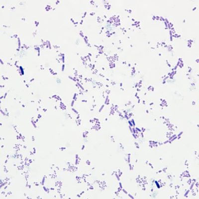
The Modified Wade-Fite Method is a staining technique used in the laboratory to detect acid-fast bacteria, particularly Mycobacterium leprae, which is responsible for causing leprosy. This staining method is a modification of the conventional Ziehl-Neelsen stain and is employed to enhance the visibility of Mycobacterium leprae in clinical specimens, such as skin biopsies.
Here’s a step-by-step guide on how to use the Mycobacterium leprae Stain Solution employing the Modified Wade-Fite Method:
Materials Required:
- Mycobacterium leprae Stain Solution (Modified Wade-Fite Method)
- Specimen slides (containing tissue sections, such as skin biopsies)
- Microscope slides
- Microscope coverslips
- Staining tray or staining rack
- Eau distillée
- Laboratory safety equipment (gloves, lab coat, etc.)
Procédure:
- Preparation of Slides:
- Prepare thin sections of the clinical specimen (par exemple., skin biopsy) and affix them onto microscope slides.
- Deparaffinization and Rehydration:
- If the specimen is paraffin-embedded, deparaffinize the slides using xylene and then rehydrate through a series of alcohol washes (par exemple., 100%, 95%, et 70% éthanol).
- Staining Procedure:
- Place the slides in a staining tray or staining rack.
- Flood the slides with the Mycobacterium leprae Stain Solution, ensuring complete coverage of the specimen.
- Incubation:
- Incubate the slides for the specified duration, typically around 15-20 minutes. The exact incubation time may vary based on the specific instructions provided with the staining solution.
- Rinçage:
- Rinse the stained slides thoroughly with distilled water to remove excess stain.
- Counterstaining (Facultatif):
- Some protocols may include a counterstaining step using a contrasting stain like methylene blue. Follow the instructions provided with the staining solution for specific details.
- Dehydration:
- Dehydrate the slides by passing them through a series of alcohol washes in ascending concentrations (par exemple., 70%, 95%, et 100% éthanol).
- Mounting:
- Apply a mounting medium to the stained slides, place a coverslip over the specimen, and allow it to dry.
- Microscopic Examination:
- Examine the stained slides under a microscope equipped with appropriate magnification. Mycobacterium leprae will appear as red-stained bacilli against a contrasting background.
- Documentation:
- Document your observations and findings for diagnostic or research purposes.
Mycobacterium leprae Stain Solution, utilizing the Modified Wade-Fite Method, a groundbreaking solution designed for accurate and efficient detection of Mycobacterium leprae in diagnostic pathology. This innovative staining method is a testament to our commitment to providing healthcare professionals with cutting-edge tools to enhance leprosy diagnosis and research.
Key Features:
- Enhanced Sensitivity and Specificity: Our Modified Wade-Fite Method ensures heightened sensitivity and specificity, allowing for the reliable identification of Mycobacterium leprae in clinical specimens. This precision is crucial for accurate diagnosis and subsequent treatment planning.
- Improved Contrast and Visibility: The stain solution is formulated to provide excellent contrast between Mycobacterium leprae and background tissue, facilitating clear visualization under microscopy. This heightened visibility aids pathologists in making confident and precise diagnostic assessments.
- Optimized for Routine Laboratory Use: We understand the importance of efficiency in the laboratory setting. Our stain solution is user-friendly and can be seamlessly incorporated into routine laboratory protocols, streamlining diagnostic processes without compromising accuracy.
- Consistent and Reliable Results: With stringent quality control measures in place, our Mycobacterium leprae Stain Solution consistently delivers reliable results. Laboratories can rely on the reproducibility of staining outcomes, ensuring confidence in diagnostic assessments.
- Versatility in Application: This stain solution is compatible with a variety of specimen types, including skin biopsies and other relevant tissues. Its versatility makes it a valuable tool for diverse laboratory settings and research applications.
- Compliance with Industry Standards: Our Mycobacterium leprae Stain Solution complies with industry standards, assuring users of its safety, efficacy, and adherence to regulatory requirements. This commitment to quality ensures the reliability of diagnostic results.
Avis des clients sur les produits Solarbio pour référence


Commentaires
Il n'y a pas encore de critiques.