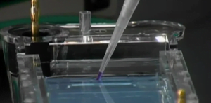
DNA electrophoresis is a common laboratory technique used to separate DNA fragments by size. It allows researchers to visualize, identify, and purify DNA samples based on their molecular weight.
Understanding the principles behind this technique, its key applications, and proper usage is essential for anyone working with DNA analysis in molecular biology, forensics, clinical diagnostics, などなど. This guide will provide a comprehensive overview of DNA electrophoresis.
What is the Principle Behind DNA Electrophoresis?
The separation of DNA fragments by electrophoresis is based on the migration of negatively charged DNA molecules through an agarose gel matrix under the influence of an electric field.
The key principles are:
- DNA molecules have a uniform negative charge due to their phosphate backbone. This means their charge-to-mass ratio is constant.
- When an electric current is applied, the negatively charged DNA molecules will be attracted towards the positive electrode.
- The agarose gel provides a sieving effect that causes smaller DNA fragments to migrate faster than larger ones.
- したがって, over time, DNA fragments become separated by size, with smaller fragments closer to the positive electrode compared to larger ones.
How Does an Agarose Gel Work?
Agarose gels are composed of agarose polymers extracted from seaweed. When agarose powder is dissolved in a buffer and allowed to set, it forms a gel containing small pores.
The features of agarose gels include:
- The concentration of agarose determines the pore size. Higher percentages make smaller pores.
- Smaller pore sizes slow migration but give better separation of small DNA fragments. Larger pores allow faster migration but lower resolution.
- Gels are submerged in a conductive buffer that maintains a constant pH during electrophoresis. Common buffers are TAE and TBE.
- An intercalating dye like ethidium bromide is added. This dye inserts between DNA bases and allows visualization of DNA bands under UV light.
What is Needed to Run DNA Electrophoresis?
The basic equipment and materials for agarose gel electrophoresis are:
- Agarose gel –通常 0.5-2% agarose concentration. Higher percentage for better separation of small fragments.
- Gel tray and comb– To cast the gel with sample wells.
- Electrophoresis tank– Houses the gel and buffer. Contains electrodes to apply current.
- Power supply –Applies a constant electric current, typically 50-150V.
- バッファ – Commonly TAE or TBE to immerse the gel. Conducts current.
- Loading dye– Contains glycerol and bromophenol blue to add density and color to DNA samples before loading into wells.
- DNA ladder– A molecular weight marker containing DNA fragments of known sizes. Used to identify sample band sizes.
- Staining dye – Ethidium bromide or alternative to visualize DNA bands under UV light.
What Are the Key Steps in DNA Electrophoresis?
The key steps in DNA electrophoresis are as follows:
- Make an agarose gel with the desired pore size and set it in the gel tray.
- Add staining dye to the gel.
- Load DNA ladder and samples mixed with loading dye into the wells.
- Immerse the gel in the electrophoresis buffer in the tank.
- Attach electrodes and apply a current to run the gel.
- Turn off the current once the dye front has migrated an appropriate distance.
- Visualize DNA bands under UV light and photograph them for analysis.
- Compare band sizes to the DNA ladder to determine fragment lengths.
- Excise DNA bands from the gel for downstream applications if needed.
What Are the Main Applications of DNA Electrophoresis?
There are many important uses of DNA electrophoresis across research, clinical diagnosis, forensics, and biotechnology:
- Confirm the success of DNA extraction or PCR amplification by visualizing DNA presence and quantity.
- Check sizes of DNA fragments after PCR, cloning, or restriction digests.
- Compare DNA profiles for identity testing in forensics.
- Detect gene insertions, deletions, or mutations associated with genetic disorders.
- Separate and purify DNA fragments of a specific size, e.g. for cloning.
- Quantify relative amounts of DNA between samples by band intensity.
- Check for DNA degradation or contaminants from diffuse or multiple bands.
- Prepare DNA for sequencing, Southern blots, or electrophoretic mobility shift assays.
What Are Some Key Considerations When Running DNA Gels?
Following are some key considerations that should be kept in mind while running DNA gels:
- Choose an agarose concentration suitable for the fragment sizes expected.
- Include a DNA ladder in at least one lane to allow band size identification.
- Mix samples thoroughly with loading dye before loading wells.
- Avoid overloading sample wells with too much DNA.
- Run gels at appropriate voltage – higher voltages can cause band distortions.
- Stop electrophoresis once the dye front has migrated an adequate distance.
- Wear protection when visualizing DNA with UV light.
- Photograph gels quickly before bands diffuse too much.
- When excising DNA bands, use a clean scalpel on a UV tray for each fragment.
結論
So, DNA electrophoresis is an essential technique that uses the migration of charged DNA molecules through an agarose gel matrix under an electrical current to separate fragments by size. Key applications include visualizing PCR products, analyzing genomic DNA, and preparative purification of DNA fragments. Careful attention must be paid to the principles and technical details when carrying out electrophoresis to obtain optimal results. With practice, DNA electrophoresis becomes a reliable workhorse technique for many molecular biology laboratories.
FAQS
Why does electrophoresis separate DNA by size?
Despite having the same charge density, the sieving effect of agarose gel pores causes DNA friction to be proportional to size – smaller fragments meet less resistance. This results in size-based separation.
How accurate is estimating fragment sizes from a DNA ladder?
Comparison to a ladder will provide an approximation of band sizes, usually within 10% 正確さ. More precise sizing requires specialized Fragment Analysis.
Why is a ladder run on the same gel as samples?
Running a DNA ladder alongside samples allows you to visualize migration in that specific gel and account for any distortions in-band migration.
Can you quantify the DNA amount directly from band intensity?
While relative intensities provide an estimate, accurate DNA quantification requires specialized instruments like spectrophotometers or fluorometers.
Why are crisp, clear bands important in DNA electrophoresis?
Sharp DNA bands indicate intact, pure DNA fragments. Diffuse bands or smears suggest degraded DNA or contaminants like leftover primers or salts that interfere.
Can electrophoresis separate DNA by sequence?
いいえ, agarose gel electrophoresis separates only by size. しかし, special techniques like DGGE can also separate based on minor sequence differences.